Date: November 22, 2023 (updated: December 3, 2023) Category: Blog, Dog health,
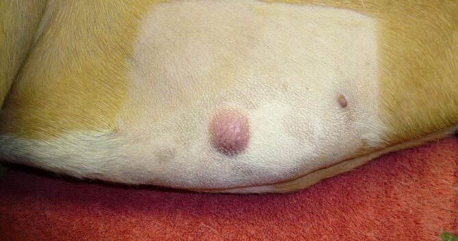
Mastocytoma is a pathological growth of mast cells, most often taking the form of a tumor occurring in the dermis or subcutaneous tissue. This type of tumor is one of the most common skin cancers in dogs, almost every fifth is a mast cell tumor. Although it has been proven that it occurs as a result of the destructive impact of substances released by mast cells, the cause of the disease has not yet been clearly established. However, it is known that the frequency of mastocytoma in dogs is influenced by breed predispositions and the age of the four-legged patient. The tumor usually appears on the dog's body, limbs or head. Since mastocytoma is often malignant and difficult to distinguish from other skin tumors, and the mere feeling of the lump does not provide clear information about its type, it is important to immediately consult a veterinarian to determine the nature of the lesion. It is worth remembering that detecting dangerous skin cancers at an early stage can significantly increase the chances of your pet's recovery.
Mastocytoma is a tumor composed of degenerated mast cells (mast cells). These cells are part of the immune system, taking over important tasks of defending the body against bacteria, allergens, toxins, viruses and other microorganisms. In response to the threat of pathogens, they can react violently, releasing substances with a strong defensive effect. One of the main substances secreted by mast cells is histamine. She is responsible, among others, for: for: itching, swelling, redness of the skin, headache, irritation of the nasal mucosa or indigestion. The release of excessive amounts of histamine and other compounds can lead to a severe allergic reaction and contribute to dangerous changes throughout the body. Mast cell tumors most often appear in the dermis or subcutaneous tissue layer.
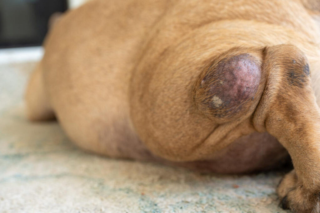
Mastocytoma in dogs is one of the most frequently diagnosed skin cancers. Basically, they can appear anywhere on the body. Most often, in the first stage, tumors are visible on the limbs, torso, head, neck or groin. Next, they may appear around the anus or on the genitals. Occasionally, they also occur on mucous membranes (e.g. oral or vaginal mucosa), ureters and spinal cord. In addition to local changes, mastocytoma may cause systemic changes, mainly affecting internal organs: liver, spleen, and gastrointestinal tract.
Division of the disease into degrees of differentiation
From the point of histological examination, mastocytoma can be divided into three degrees of differentiation:
Grade I – the least malignant, with well-differentiated tumors, slightly invasive (slow growing) and low metastasis rate. Overall, complete resection of such tumors leads to complete cure in 85% of cases.
Grade II – mastocytoma of cells of intermediate histological grade, most often located in the subcutaneous tissue. The disease requires additional tests to determine the scale of mastocytoma aggression, including: the Ki 67 antigen (a core protein created by dividing mast cells) and the c-kit mutation (a receptor responsible for regulating the growth, differentiation and activation of mast cells ). In this case, complete removal of the tumor in healthy tissue, provided that no other organs (especially lymph nodes) are affected, gives a good prognosis.
Grade III – mastocytoma with the highest degree of malignancy, giving the worst prognosis. The tumors are large, infiltrate the subcutaneous tissue and epidermis, and their surface is usually ulcerated. Unfortunately, the disease is extremely aggressive and quickly spreads throughout the body. It has the shortest survival time after surgery, unlike four-legged patients with lower-grade tumors. It is worth remembering that none of the individual factors clearly determines the prognosis; the entire clinical picture of the sick dog should be taken into account.
A tumor visible on your pet's body is a clear sign that you need to go to the vet to have it checked out!
What causes mastocytoma in dogs? Overview of risk factors
The causes and risk factors for the development of mast cell tumors are not yet sufficiently researched. Genetic variations are believed to play an important role and, in combination with environmental factors, lead to malignant degeneration of mast cells. A mutation in the c-kit gene was found in approximately 30% of four-legged patients with mastocytoma. Some dog breeds are also indicated, such as: boxer, golden retriever, beagle, labrador, rhodesian ridgeback, hound dog, pug, fox terrier, bulldog, Weimaraner, which affects this disease more often. Fortunately, recent reports show that the vast majority of these cases involve a less malignant type of mastocytoma. The tumors are well differentiated, have a benign clinical picture and have a favorable prognosis. The situation of representatives of the breed is different shar-pei, in which mastocytomas are often poorly differentiated, have a high degree of malignancy, and are much more aggressive.
In addition to genetic predisposition, statistics indicate that mastocytomas are diagnosed more often in older dogs, starting from the age of eight. However, it must be emphasized that mastocytoma can occur in dogs of any age. It is diagnosed even in young dogs as young as four months old. We are encouraged by studies confirming a reduced risk of mastocytoma in some breeds. Among them is: Shih Tzu, Maltese, yorkshire terrier, chihuahua, dachshund, miniature poodle.
Symptoms of mastocytoma in dogs
Mastocytoma manifests itself with various skin lesions. In the first type, most tumors occur in the form of cutaneous or subcutaneous lesions with a diameter of up to 4 cm. They are clearly demarcated from the surrounding tissues and grow quite slowly. The second type consists of poorly defined tumors, which take the form of diffuse, swollen skin lesions with a soft structure and poorly defined borders. This type of mastocytoma is characterized by rapid growth and a tendency to ulcerate. Some tumors do not change their size for many weeks, but after some time they begin to change their appearance and spread very quickly. Other mastocytomas are aggressive from the very beginning.
In the case of disseminated or systemic mastocytoma, the most common symptoms are: generalized lymph node enlargement, liver enlargement, and spleen enlargement. These symptoms are most often accompanied by: apathy, weight loss, decreased appetite, vomiting, and diarrhea.
Unfortunately, the symptoms associated with the primary tumor and the secondary tumor are often accompanied by clinical symptoms resulting from the destructive effects of substances released by mast cells. As a consequence, metastases occur to internal organs, including: in nearby lymph nodes, in the spleen and liver, and less often in the lungs. Stomach ulcers and intestinal ulcers appear, usually in the pyloric part of the stomach. The disease causes abdominal pain, diarrhea, decreased appetite, bloody vomiting and tarry stools. Occasionally
the gastrointestinal wall is perforated.
Moreover, due to the release of significant amounts of heparin by mast cells, problems with blood clotting and wound healing occur. In case of local bleeding, it may be difficult to stop the blood. Another effect of the destructive impact of mastocytoma is a whole range of allergic symptoms with varying degrees of severity: from a local skin reaction (swelling, itching, hives) to anaphylactic shock, which threatens the pet's life.
What diagnostic tests does the vet perform?
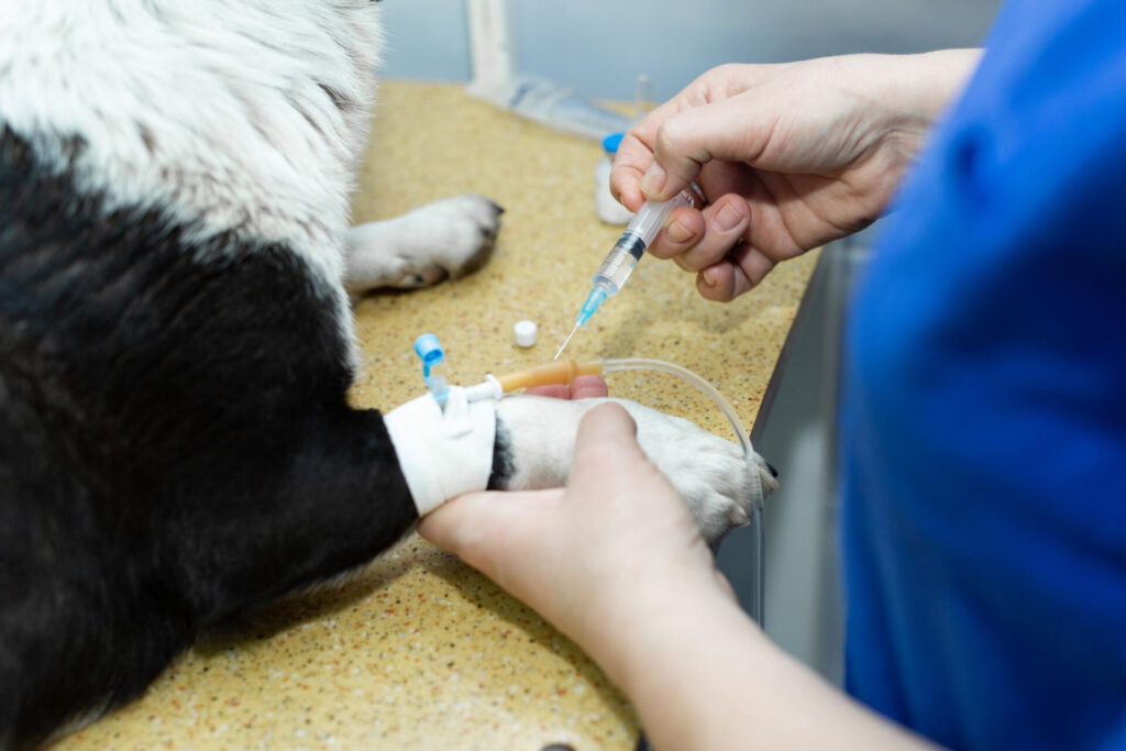
When diagnosing mastocytoma, in addition to a general examination by a veterinarian, a fine-needle aspiration biopsy with puncture of the nodule or a biopsy is performed. Histopathological examination of the collected material makes it possible to determine the degree of malignancy of the tumor. Thanks to this, it is possible to determine the optimal therapeutic procedure, plan appropriate treatments, form of pharmacological treatment and outline the prognosis. In the initial diagnosis of mastocytoma, a biochemical blood test and a complete blood count as well as a general urine test are also used. In-depth diagnosis (in the case of metastases) includes: X-ray of internal organs, abdominal ultrasound, cytological examination of the bone marrow and surrounding lymph nodes, as well as cytological examination of a sample from the liver or spleen (optional).
Treatment strategies for dogs with mastocytoma
The selection of the treatment method depends primarily on the stage of the disease, the location of the tumor(s), the histopathological grade of the lesion and the general health condition of the dog. Grade I and II mast cell tumors most often qualify for complete removal during surgery. Complete resection of the mastocytoma with a 2 cm margin on each side of the lesion and removal of 1 fascia below the tumor is an important factor in the prognosis. During surgery, the surgeon strives to obtain the cleanest cutting margins possible.
In some cases, the therapeutic process after complete resection of mastocytoma is additionally enhanced with chemotherapy or radiation. When it comes to poorly defined tumors occurring on the limbs, in most cases a radical surgical procedure is recommended, i.e. amputation of the affected limb. When the tumor cannot be removed due to its location or it is a grade III mastocytoma, adjuvant treatment is introduced, i.e. supplementing surgical treatment with radiotherapy and chemotherapy. If the mastocytoma has spread, chemotherapy may improve the dog's functioning and prolong its life. Similarly, radiotherapy, which reduces the size of individual tumors, can improve the quality of life of a four-legged patient. It has also been shown that drugs such as prednisone can be successfully used before surgery to reduce the size of mastocytoma, which in turn allows surgery to be performed. In the treatment of dogs, after removal of a poorly differentiated mast cell tumor, vinblastine, used together with prednisone, is effective.
Chemotherapy is often necessary to treat mastocytoma.
Life with a dog suffering from mastocytoma
Cancer is a huge challenge for your four-legged friend's body. A dog affected by mastocytoma must cope with weakness, malaise and pain. Cancer cells mainly use carbohydrates as an energy source. They also deprive the body of important nutrients and energy. The pet loses weight, becomes apathetic, weak and exhausted. Because the disease has a significant impact on the proper functioning of the entire body, depending on the course of the disease, it changes the body's demand for particular nutrients to varying degrees. And it is in this area that we can effectively support the therapeutic process and improve the quality of life of our sick pet.
By following a special diet, we can, to some extent, limit the negative consequences of mastocytoma, improve the functioning of the body and prevent its destruction. When preparing meals for a patient, remember about the appropriate proportions of individual nutritional ingredients. You need to provide a high-energy menu, rich in fats and proteins, and eliminate carbohydrates from your diet. The share of protein in portions should be 35-50%, fat 50-60%. It is worth enriching your diet with polyunsaturated fatty acids from the omega 3 group and glutamine, arginine, leucine and valine. Mineral and vitamin supplements will also be important - especially with selenium and vitamins A, C, E. You can use ready-made special food, including: for dogs: convalescents, fussy, sensitive or with digestive tract disorders.
In addition to an appropriate diet, a very important element during therapy and convalescence is providing proper care for the patient at home. All recommendations of the veterinarian should be followed, in particular the timing and dosage of medications and the selection of the level of physical activity. You should carefully monitor the condition of your pet's skin, regularly monitor the site of mastocytoma removal and attend scheduled check-ups. We also try to provide your pet with peace and a sense of security.
How to best support your pet when sick?
Unfortunately, mastocytomas, i.e. mast cell tumors, do not have a characteristic shape. They come in many shapes and sizes and in any part of the body. They can be large, small, hard, soft, or even ulcerated. For this reason, it is important to regularly examine your pet during check-up visits to the vet and to be constantly vigilant in this matter on a daily basis. Additionally, given that virtually any skin lump can be a mastocytoma, it is important to take your dog to a veterinarian as soon as possible if you notice any skin lesions.
Please remember that mastocytomas are among the most common skin cancers in dogs. The prognosis depends on the stage of the cancer and the location of the tumor. Catching a low-grade lesion in time increases the chance of your four-legged friend's complete recovery. Therefore, it is worth emphasizing once again that regular examination for new skin lesions is the most important preventive measure in the prevention of mastocytoma.
Behavior that deviates from normal or is specific to the dog's breed, age or sex is called behavioral problems. The causes of such problems may be genetic, organic or due to environmental influences. When the dog's guardian is unable to cope with the pet's complicated behavioral disorders on his own. They can use the help of a dog behaviorist who, by understanding the cause and function of problematic behaviors, helps alleviate or eliminate them.
The German director, actor and humorist Vicco von Bülow, also known as Loriot, not without reason claimed that life without a pug is possible, but completely meaningless. After all, he is a charming companion with a charming, cheerful and friendly disposition, whose large eyes and wrinkled face are hard to resist.
Dogs are born blind and it can take up to two weeks before they open their eyes for the first time. And although their sense of vision improves significantly over the following months, it is believed that in the world of colors, it is humans who have the advantage. Is it right? There is a big difference between how a dog sees and how we see the world. For a long time, dogs were thought to be colorblind. In fact, they cannot see a specific color spectrum as clearly as humans. However, in return, they notice things that remain hidden from our sense of sight.


